A new MRI neuroimaging facility launched at Pennovation Works earlier this year with the goal of helping Penn researchers understand the relationship between the human brain and behavior.
The space, known as the MindCORE MRI Neuroimaging Facility, hosted an open house event on Friday to showcase its new MRI scanner. The facility aims to further research from various Penn neuroscience, psychology, and cognitive science labs that utilize multimodal imaging techniques.
“The goal here is to provide access to the resource for a large community of people [within the University]," said MindCORE's director Joseph W. Kable in Omnia.
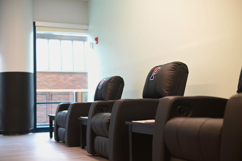
The space also includes three behavioral testing rooms, conference rooms, and office spaces for research personnel.
MindCORE is Penn's center for the integrative study of the mind, bringing together faculty across various departments. The University established it under the "Mapping the Mind" initiative, part of the School of Arts and Sciences' strategic plan outlined by SAS Dean Steven J. Fluharty.
While there are many resources for MRI research on Penn's campus, they are shared across different departments and primarily housed in Penn Radiology, said Executive Director of the Neuroimaging Facility Brock Kirwan in an interview with The Daily Pennsylvanian.
“The impetus for this facility was people doing cognitive neuroscience who are really interested in a particular type of MRI scanning: functional MRI scanning,” Kirwan said.
Standard MRI scans are used to produce detailed anatomical images of soft tissues in the body. The MRI creates a powerful magnetic field that interacts with protons found in the water molecules that makes up living tissues. In scans of a shoulder or an ACL tear, for example, healthy and injured tissue show up differently due to the differences in their magnetic properties.
Functional MRI scans focus solely on the brain and measures the brain's functional activity in addition to producing an image. An fMRI does this by tracking oxygen levels throughout the brain.
While the MRI scanner in the Neuroimaging Facility is not the strongest MRI scanner on campus, it is the one best equipped for specifically studying cognitive processes.
Kirwin, whose research background is in memory, described potential uses for the MRI scanner in memory studies.
“We would have you come into the facility, show you some stimuli in one of [the testing] rooms, and test your memory for these a little bit later,” Kirwan said.
Participants may first be asked to answer a question about themselves or objects in their vicinity. They would then enter the MRI scanner while researchers administer a memory test. The machine could track differences in brain activity when participants look at images of the objects they were just exposed to in the testing rooms compared to new stimuli.
The last piece of equipment that the facility is waiting on is an MRI-compatible monitor that will sit outside of the scanner. The monitor will be set up in conjunction with a mirror that allows participants to see stimulus images on the monitor while lying on the scanner bed.
Participants will also be given a button box or joystick to allow them to make responses while in the machine. They can indicate their answers to yes or no questions or indicate if an image is similar or different to an image they have seen before.
Additionally, researchers may manipulate some aspects of the stimulus and try to trick participants’ brains into “false-alarming,” which the scanner will be able to detect and record different forms of brain activity, Kirwan said.
This facility will welcome all types of academic study, including serving as an undergraduate classroom.
Kirwan plans to teach a class next semester on fMRI methods, in which students will learn the physics behind how the machine works, and how to design an fMRI experiment. He plans to collect data from the class and help students learn to run their own experiments.
Correction: A previous version of this article erroneously stated that Fluharty was the former College of Arts and Sciences dean. The article has been updated to clarify that he is the current dean of the School of Arts and Sciences. The DP regrets this error.
The Daily Pennsylvanian is an independent, student-run newspaper. Please consider making a donation to support the coverage that shapes the University. Your generosity ensures a future of strong journalism at Penn.
Donate


